Hip & Knee Chart * Shows hip and knee general anatomy * Also illustrates posterior anterior and lateral view of the hip joint as well as tibial plateau * Shows total hip replacement hip fracture repair and blood supply to the head of the femur * Shows various kinds of meniscus and ligament tears * Provides anterolateral posteromedial and posterior view of the knee * Shows arthroscopy and total knee replacement * Colorful laminated and detailed * 20 W x 26 *
This chart illustrates how ones posture changes due to the different types of spinal disorders * The chart shows tumors on the spinal column ilium sacrum and spinal cord arthritis of the hip herniated disc fractures of the vertebrae and sacrum and the effects of osteoporosis on bones * Laminated * 20 x 26
Ligaments of the Joints * Shows location of various joints provides anterior and posterior views of the left shoulder right hip right knee and left elbow * Also illustrates lateral and medial views of the left elbow and ankle joint * Shows superficial volar deep dorsal and superfical dorsal views of the left wrist *
Lymphatic System * Classic illustrations by Peter Bachin. Shows system throughout the body * Illustrates internal iliac lymph node and lymph vessels & lymph nodes of the stomach pancreas spleen and biliary tract. Also shows lymph vessels & lymph nodes of the small intestine and lymph vessels & lymph nodes of the inguinal region pelvis and the posterior wall of the abdomen * Colorful laminated and detailed * 20 W x 26 * Shipping Carton Size: 41 L x 8 W x 4 H
Male Reproductive * Provides anterior and posterior view of the system * Shows the pelvic organ (oblique section) and cross-section of the penis * Illustrates the prostate perineum spermatogenesis testis and vasculature & innervation * Colorful laminated and detailed * 20 W x 26 *
Muscular System * Classic illustrations by Peter Bachin * Shows anterior and posterior views of the muscular system * Also illustrates right half of the diaphragm muscles of the posterior abdominal wall and muscles of the right and right foot * Colorful laminated and detailed * 20 W x 26 *
Nervous System * Classic illustrations by Peter Bachin * Illustrates nerves in the body brain midbrain medulla oblongata and spinal cord * Also shows spinal meninges intercostal nerves and sagittal section of female pelvis * Colorful laminated and detailed * 20 W x 26 *
Great for busy therapy sessions * Keeps ice and ice packs conveniently and readily available * Expanded polymer foam is virtually indestructible * Withstands temperatures between -321 - 200F (-196 to 93C) * Constructed of expanded polymer foam that keeps ice from melting longer than traditional ice pails * The 4 liter capacity is ideal for keeping small readymade ice packs close at hand * Lightweight and easy to transport the ice bucket is virtually indestructible impervious to moisture and odor and will not sweat * 8 1/2 opening at top x 6 3/5 high (21.5 x 17 cm) bottom tapers to 6 3/10 (16 cm) at the base *
Shoulder & Elbow Chart * Illustrates general shoulder and elbow anatomy * Shows anterior posterior lateral and superior view of the shoulder * Also shows the socket of shoulder joint anterior and disclocation of humerus * Illustrates impingement syndrome and acromioplasty * Shows sagittal view of the elbow as well as supination pronation and superior views of extension and flexion. Illustrates elbow fractures and tennis elbow *
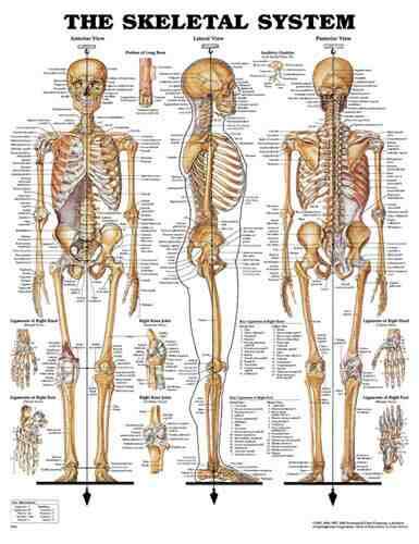
Skeletal System * Classic illustrations by Peter Bachin * Shows anterior lateral and posterior views of the skeletal system * Also illustrates portion of long bone auditory ossicles ligaments of the right hand (dorsal and palmar views) ligaments of the right foot (dorsal and plantar view) and the right knee joint (anterior and posterior views) * Colorful laminated and detailed * 20 W x 26 *
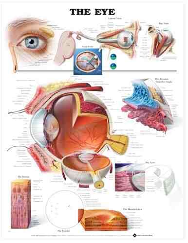
Shows cross section of the eye * Provides lateral and top view of the eye and shows the visual field * Illustrates anterior chamber angle lens retina fundus and the macula lutea * 20 W x 26 H *
Vascular System * Classic illustrations by Peter Bachin * Shows system on body * Illustrates heart (right interior left interior and posterior views) heart in systole female pelvis base of the brain and branches of abdominal aorta and portal vein * Colorful laminated and detailed * 20 W x 26 *
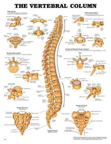
Vertebral Column Chart * Highly detailed life-size model shows all significant features for each vertebra including vertebral body spinous and transverse processes vertebral notch and spinal canal * Features complete pelvis sacrum occipital bone vertebral artery and nerve branches * Herniated disc between the 3rd and 4th lumbar vertebrae * Ideal teaching model for students for patient education by chiropractors orthopedic surgeons and other medical professionals and for company health programs on lifting and bending * 2-part stand included. Size: Spine 29 tall stand 34 1/2 *